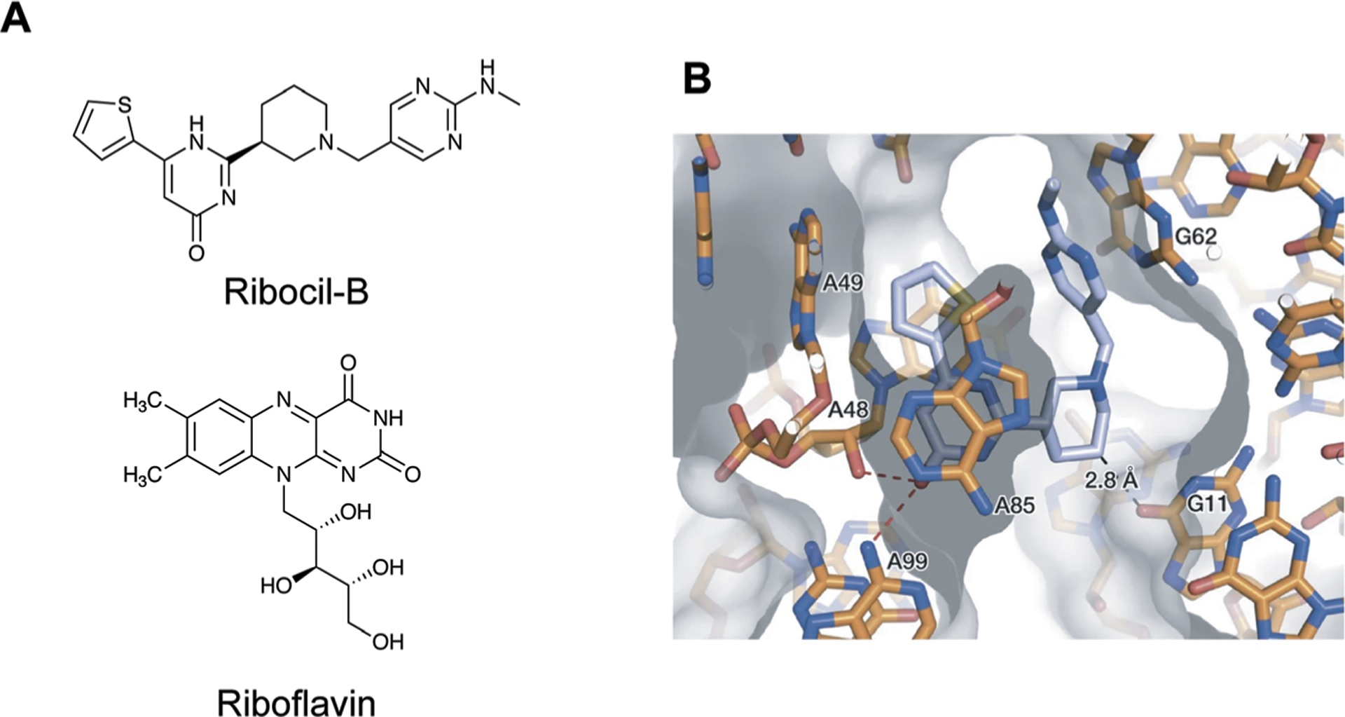
Fig. 13.

Small molecule targeting of the riboflavin riboswitch. (A) The structures of Ribocil-B and Riboflavin. (B) Crystal structure of Ribocil-B (blue sticks) in complex with riboflavin riboswitch. Ribocil binding is stabilized by stacking interactions with A48 and A85 and hydrogen bonding between the oxygen and A48 and A99. Additional stacking interactions and a methyl hydrogen bond are observed on the other face of the molecule. Adapted from ref. 13 with permission from Springer Nature, Copyright 2015.
