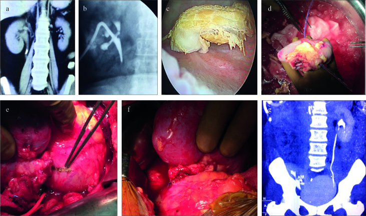
Figure 2. a–g.
(a) Preoperative computed tomography (CT). (b) Postavulsion nephrostogram. (c) Avulsed ureter segment seen inside the urinary bladder. (d) Retrieved kidney with feeding tube from nephrostomy site coming out from the ureter. (e) Bladder marked for ureteroneocystostomy. (f) Completed ureteroneocystostomy. (g) Postoperative CT scan with autotransplanted kidney in the right iliac fossa region

