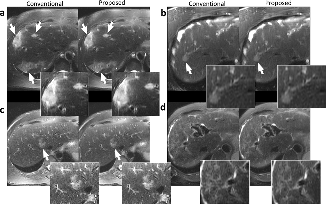
Figure 4.
Example images of the liver using conventional reconstruction (left) and the proposed data-driven reconstruction (right) on a 73-year-old female patient with focal liver lesions (a), a 67-year-old female patient with a focal liver lesion (b), a 52-year-old female patient with a focal liver lesion (c), and a 58-year-old female patient with cirrhosis (d). Regions with potential lesions are indicated by white arrows. The proposed method and the conventional reconstruction achieved comparable structural delineation of the lesions. Less perceived noise can be observed in (a) and (c) in the proposed reconstructions.

