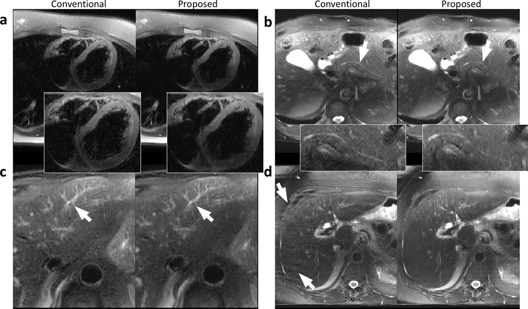
Figure 6.
Example images containing other regions of interest or artifacts using the conventional reconstruction (left) and the proposed data-driven reconstruction (right) on a 25-year-old female patient (a), a 66-year-old male patient (b), a 73-year-old male patient (c), and a 64-year-old female patient (d). Comparable sharpness of the heart was achieved using the proposed method (a) with reduced perceived noise. The pancreatic duct was visible in both conventional and proposed reconstructions (b) with reduced perceived noise using the proposed method. Small structures in the liver can be reconstructed and share similar quality with both methods (c). The data-driven approach reduces the phase-wrapping artifacts occurred in the conventional reconstruction in (d).

