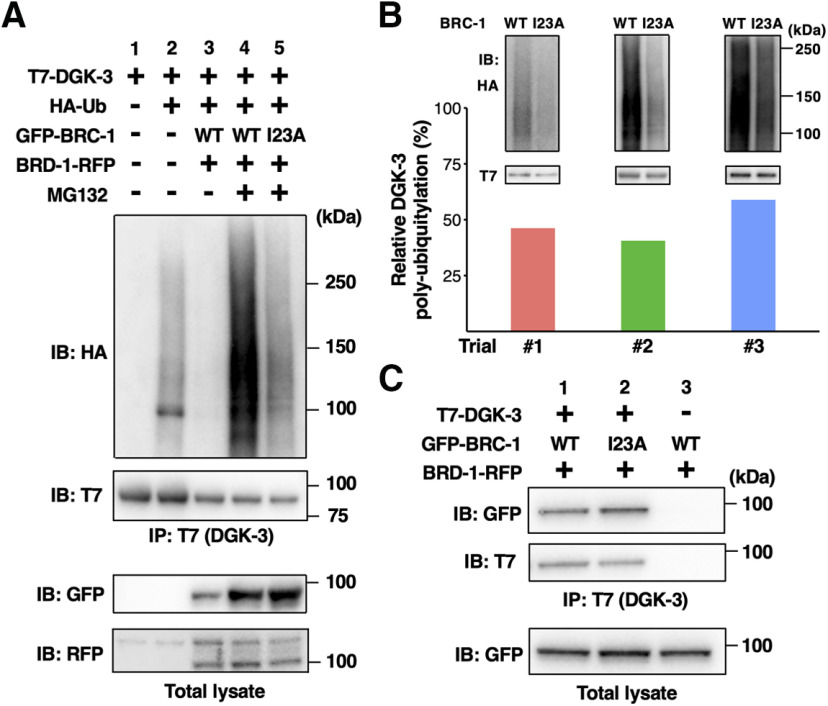
Figure 7.
BRC-1–BRD-1 mediates poly-ubiquitylation of DGK-3. A, Poly-ubiquitylation of DGK-3 by BRC-1–BRD-1. COS-7 cells were transfected with T7-DGK-3, HA-Ub, GFP-BRC-1, and BRD-1-RFP, as indicated. Cells were incubated with or without MG132. Cell lysates were immunoprecipitated (IP) with anti-T7 antibody and immunoblotted (IB) with anti-HA and anti-T7 antibodies. Total lysates were analyzed by IB with anti-GFP and anti-RFP antibodies. The experiment was done in triplicate with similar results, shown here for trial #2. B, Comparison of DGK-3 poly-ubiquitylation levels. The DGK-3 poly-ubiquitylation experiment was performed three times and each bar represents the result of each trial (#1–#3). Data represent the percentage of normalized poly-ubiquitylated DGK-3 in lane 5 relative to that found in lane 4. The blots in lanes 4 and 5 of A from three trials are shown in the upper part. C, Interaction of DGK-3 with BRC-1. COS-7 cells were co-transfected with T7-DGK-3, GFP-BRC-1, and BRD-1-RFP, as indicated. Cells were then incubated with MG132. Cell lysates were immunoprecipitated (IP) with anti-T7 antibody and immunoblotted (IB) with anti-GFP and anti-T7 antibodies. Total lysates were analyzed by IB with anti-GFP antibody.

