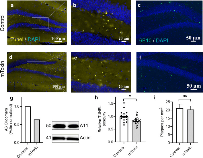
Fig. 6.
a, d TUNEL staining in the hippocampi of 3×Tg mice (40×magnification). b, e Dentate gyri digitally zoomed-in. c, f 6E10 staining of 3×Tg mice coronal sections detects Aβ plaques of various dimensions (green objects). g Kinetworks™ Custom Multi-Antibody screen 1.0 western blot of pooled lysates (four animals each group). h Relative TUNEL-positive surface area (unpaired two-tailed t-test, *p<0.05). i Amyloid-β plaques’ density in the hippocampi of 3×Tg mice (t-test) n=16, four mice

