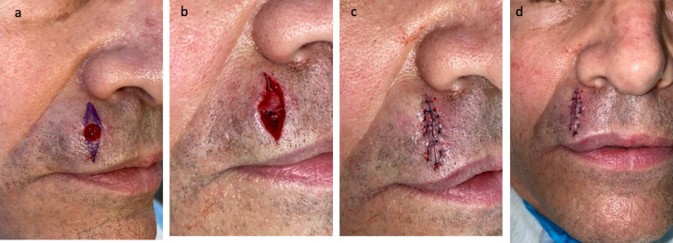
Fig. 3.
Excision of red dot basal cell carcinoma and closure of the surgical wound on the right side of the upper lip of a 65-year-old male. Correlation of the clinical presentation and the pathologic findings of the lesion on the right side of the upper lip of the 65-year-old male was a red dot basal cell carcinoma (with superficial and nodular tumor aggregates). The tumor was excised using the Mohs technique. Curettage of the site prior to the initial incision was unremarkable. One stage was required for cancer removal. The postoperative defect was 8 × 9 mm; the purple markings show the planned excisions of the standing cones to convert the oval wound into an ellipse (a). The final defect (b) was closed with a complex linear repair using a synthetic absorbable monofilament 5–0 polydioxanone (PDS) suture for closing the dermis and a synthetic nonabsorbable monofilament 6–0 polypropylene (prolene) suture for bring the epidermal edges together; side (c) and frontal (d) views show the 2.8 cm linear closure on the right side of the upper lip after completion of suturing. There has been no recurrence 3 months after surgery

