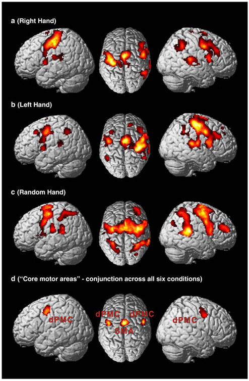
Fig. 2.
(a) Pattern of activation for right-hand movements consisting of left primary motor and somatosensory cortex, thalamus and insula as well as bilateral secondary somatosensory cortex, basal ganglia, supplementary motor area, dorsal and ventral premotor cortices. Furthermore the right temporo-parietal junction and middle frontal gyrus were activated. For left hand movements we found a mainly mirror-reversed pattern of activity (b). Random hand movements (c) feature bilateral activation of the primary sensory-motor and cortex, putamen, pre-supplementary and supplementary motor area, ventral premotor cortex, cingulate motor cortex as well as the intraparietal sulcus and temporo-parietal junction. An additional bilateral cluster is localised on the superior frontal gyrus anterior to BA 6. Right-hemispheric activation was observed in pars opercularis of the right inferior frontal gyrus (BA 44). (d) Areas which have been constantly active throughout all condition and hence represent the “core motor areas”. Activity was found bilateral in the dorsal premotor cortex, the mesial aspect of the frontal lobe (SMA) and the putamen.

