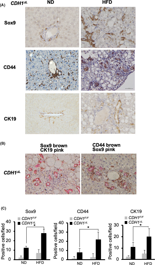
FIGURE 2.

NAFLD increased Sox9‐, CD44‐, and CK19‐positive cells in E‐cadherin‐deleted liver. A, Immunostaining of Sox9, CD44, and CK19 in CDH1ΔL ND and HFD mice (×200 magnification). B, Double immunostaining of Sox9 (brown)/CK19 (pink) (left panel) and CD44 8brown)/Sox9 (pink) (right panel) in CDH1ΔL HFD mice (×200 magnification). C, Quantification of Sox9, CD44, and CK19 cells in CDH1ΔL (n = 5) and CDH1F/F (n = 5) mice. Positive cell numbers per field. *P < .05
