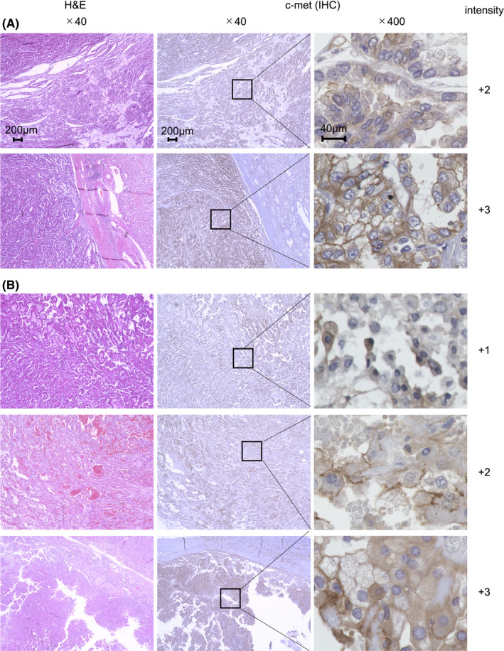
FIGURE 1.

Expression of c‐met in clinical samples of PRCC. H&E staining and immunohistochemistry (IHC) for c‐met were conducted on clinical specimens of type 1 (A) and type 2 (B) PRCC. In IHC analysis, rabbit anti‐human c‐met mAb were employed for primary staining. Microscopic examination of H&E and IHC staining samples were conducted at ×40 or ×400 magnification. The values of c‐met intensity were indicated. Representative images are displayed
