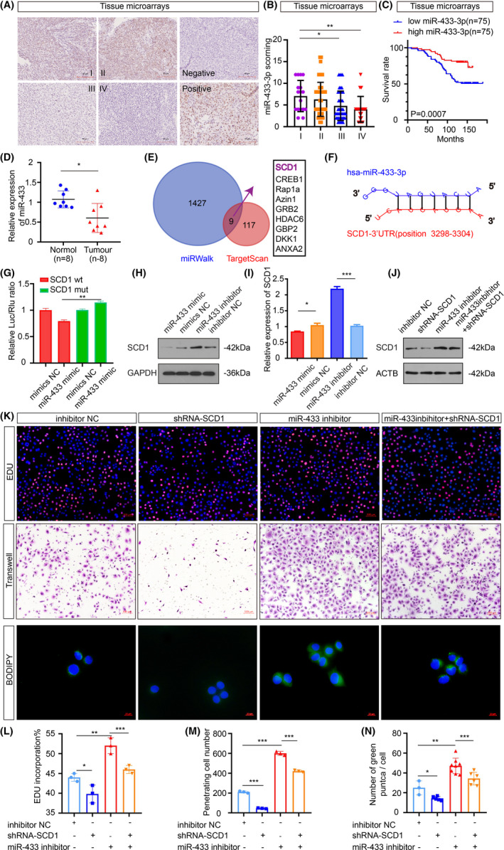
FIGURE 4.

Stearoyl‐CoA desaturase 1 is a direct target gene of miR‐433‐3p. A, Representative miR‐433‐3p in situ hybridization staining of NPC tissue microarrays. Scale bar: 90 μm. B, Statistical comparison of miR‐433‐3p expression across clinical stages using one‐way ANOVA. C, Patients were classified into high and low expression groups according to the median value of the miRNA expression. Kaplan‐Meier analysis was used to compare the overall survival. D, The relative expression of miR‐433‐3p in normal and NPC samples (n = 8 per group) was measured by qRT‐PCR. E, Venn diagram depicting predicted miR‐433‐3p targets. F, Schematic of predicted miR‐433‐3p binding sequences in the 3′‐UTR of SCD1. G, Luciferase reporter assay was conducted to confirm the relative luciferase activities of wild and mut SCD1 reporters. H, I, The protein and mRNA expression of SCD1 when miR‐433‐3p expression was increased or decreased. J, Rescue experiments with Western blot of changes in SCD1 protein levels induced by miR‐433‐3p suppression and SCD1 knockdown. K, CNE2 cells were treated with different plasmids for 48 h. Proliferation, migration, and lipid synthesis were determined by EdU (L), Transwell (M), and BODIPY staining (N). *P < 0.05; **P < 0.01; ***P < 0.001
