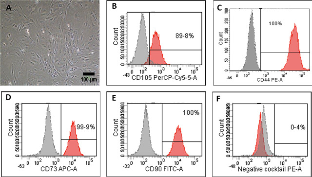
Figure 1. Characterization and differentiation potential of ADSCs.

Characterization and differentiation potential of ADSCs. (A) Primary ADSCs morphology after 14 days in culture under the inverted phase contrast microscope. Scale bar = 100 µm. (B–F) Flow cytometry staining of ADSC markers. Cells showed positive staining for mesenchymal stem cells markers CD-44, CD-105, CD-73, CD-90 and negative for CD-34, CD-11b, CD-19, CD-45 and HLA-DR in the negative cocktail.
