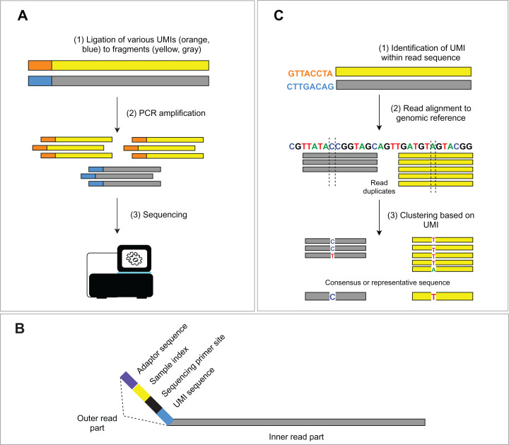
Figure 2. Schema of UMIs experimental and bioinformatic processing.
(A) During NGS library preparation, UMIs are ligated to DNA fragments, followed by PCR amplification and sequencing. (B) Structure of the outer part of a read is depicted. (C) Bioinformatic read deduplication and error correction using UMI identification and read clustering. Depending on the selected tool, deduplication results either in a consensus read creation or the selection of the most representative read.

