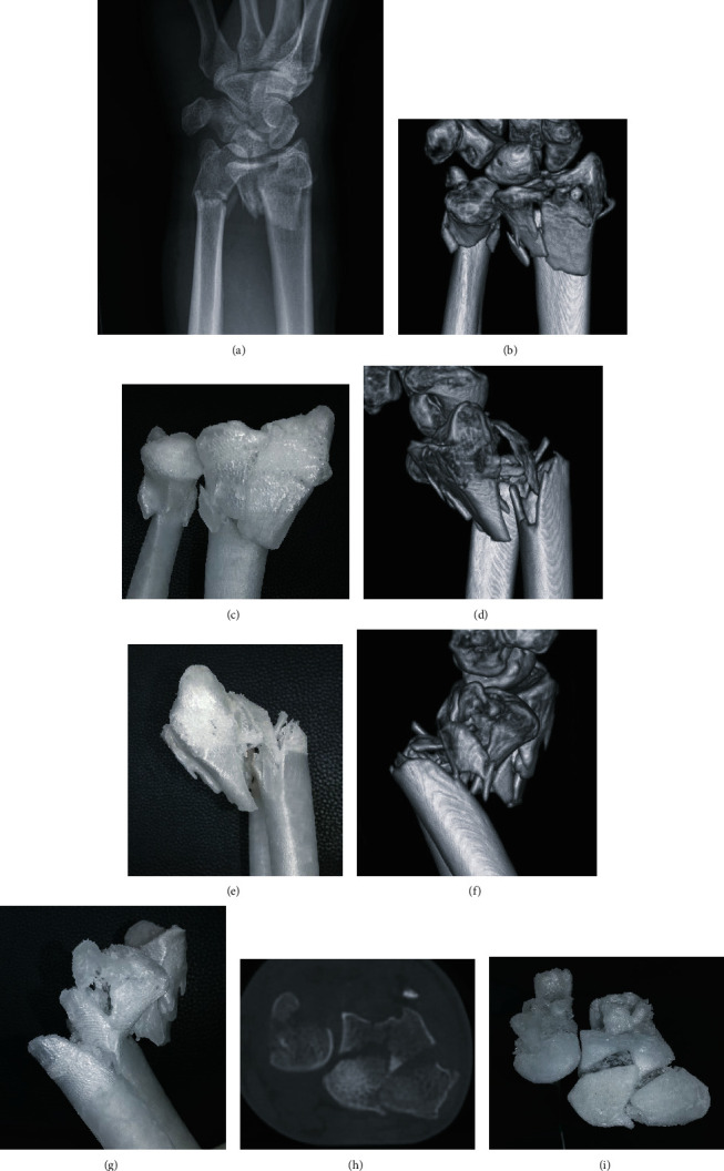
Figure 3.

(a) Dorsopalmar radiographic view of a displaced, multifragmentary, intra-articular fracture of the distal radius, and ulna of a 45-year-old female. (b, c) Coronar view of the 3D-CT reconstruction and the equivalent view on the 3D-printed Polypropylene-model. (d, e) Sagittal view (radial aspect) of the 3D-CT reconstruction and the equivalent view on the 3D-printed Polypropylene-model. (f, g) Sagittal view (ulnar aspect) of the 3D-CT reconstruction and the equivalent view on the 3D-printed Polypropylene-model. (h, i) Axial CT-projection and the equivalent view on the 3D-printed Polypropylene-model allowing an overview on the intra-articular key fragments of the distal radius fracture.
