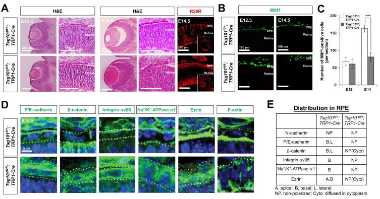
Fig. 3. Tsg101-deficent embryonic RPE cells failed to form a monolayer structure.
(A) Morphologies of the embryonic mouse eyes were examined by H&E staining of eye sections of Tsg101 fl/+ ;TRP1-Cre and Tsg101 fl/fl ;TRP1-Cre littermate mice at E12.5 and E14.5. The Cre-affected cells in E14.5 mouse eye sections was visualized by immunostaining of β-galactosidase expressed from R26R Cre reporter alleles. (B) Distribution of RPE in the littermate mouse eyes was examined by immunostaining of Mitf1. (C) The numbers of Mitf1-positive RPE in the mouse eyes were quantified and shown in the graph. Error bars denote SD (n = 4 from 4 independent litters). ***P < 0.001. (D and E) Distribution of polarity marker proteins in mouse RPE of E14.5 Tsg101 fl/+ ;TRP1-Cre and Tsg101 fl/fl ;TRP1-Cre littermate mice was investigated by immunostaining (D) and the results are summarized in (E). Dash lines in (D) indicate basal (top) and apical (bottom) margins of RPE. Blue fluorescence signals are the nuclei stained by Hoe.

