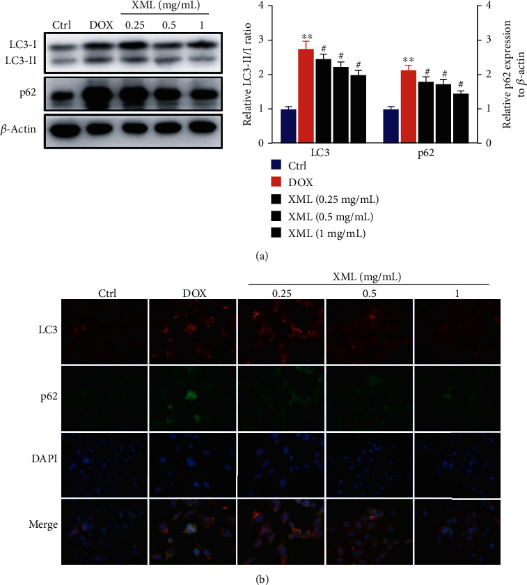
Figure 3.

XML ameliorates autophagy flux block in DOX-induced H9c2 cells. (a) Western blotting of LC3 and p62 was represented with the LC3-II/I ratio and normalization to β-actin, respectively. (b) Representative immunofluorescence images of LC3 (red), p62 (green), and DAPI (blue). H9c2 cells were treated with 5 μmol/L DOX for 24 h only or preincubated with 0.25, 0.5, and 1 mg/mL XML for 48 h. Data are representative of at least three independent experiments (mean ± SD). Statistical significance was assigned as ∗P < 0.05 and ∗∗P < 0.01 compared with control and #P < 0.05 compared with DOX. P values were determined using the independent one-way ANOVA and the Tukey-Kramer test. XML: Xinmailong treatment; DOX: doxorubicin.
