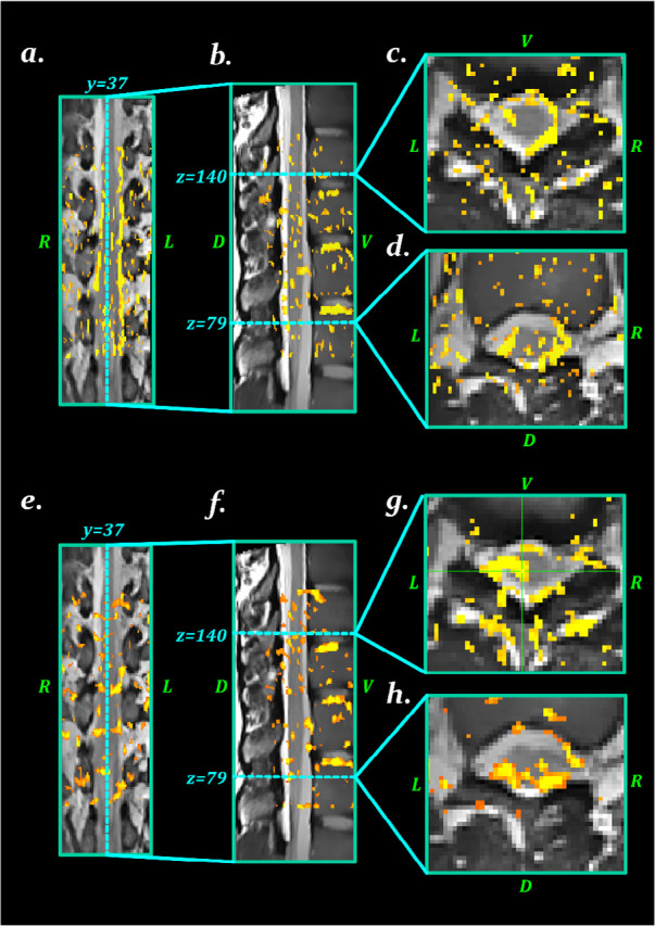
Figure 7.

The influence of noise correction methods on the activation maps
A–D: The activation maps with no noise correction; E–H: The activations map after cardiac and respiration noise correction; This figure illustrates that physiological noise correction decreases the active voxels in the CSF (false positives) and increases active voxels in the spinal cord.
