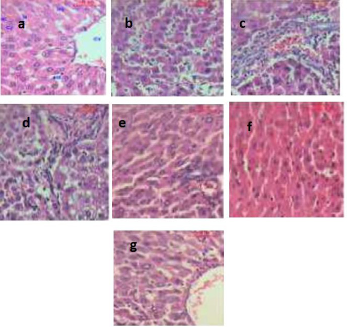
Figure 8.
It shows photomicrographs (H&E X400) of various liver tissues from control and experimental animals. (a) shows the architecture of a hepatic lobule of control rats. The central vein (CV) lies at the center of the lobule surrounded by the hepatocytes (HC) with strongly eosinophilic granulated cytoplasm (CY) and distinct nuclei (N). Between the strands of hepatocytes, the hepatic sinusoids are shown (HS), (b) shows the area of the aberrant hepatocellular phenotype of DEN group with variation in nuclear size, hyperchromatism, and irregular sinusoids, (c) shows abnormal hepatocellular histology of DEN group with prominent hyperbasophilic preneoplastic focal lesions and eosinophilic clear cell foci, (d) shows trabeculae of hepatocellular carcinoma in DEN group consists of highly pleomorphic tumor cells and degenerated tumor cells, (e) shows the portal lobules of DEN + LCB group that appear more or less like the control one. Notice the activated Kuppfer'cells, (f) shows hepatic lobule of DEN + FnC60 group resembling control one, (g) shows many of the hepatocytes in DEN + LCB + FnC60 group appear more or less like normal

