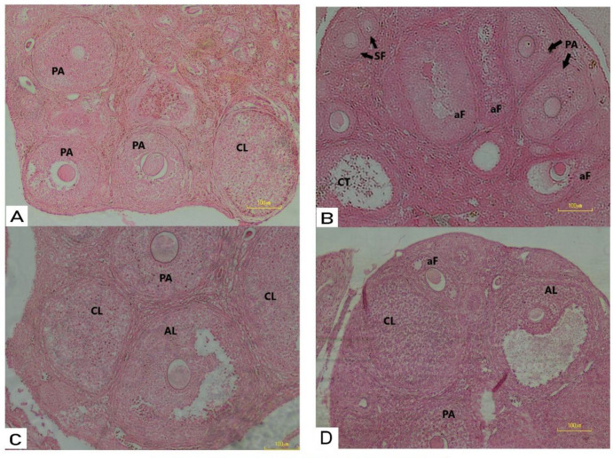
Figure 1.
(A) Histological analysis of mice ovaries Control mice ovary showing normal follicles and corpus luteum; (B) PCOS mice ovary treated with estradiol valerate showing degeneration in follicles at development stages (atresia); (C) Photomicrograph of mice ovary treated with E.V plus CC showing more active ovary than PCOS with follicles as well as corpora lutei, preantral follicle and antral follicle; (D) Mice ovary treated with E.V plus metformin showing follicles in different developmental stages, as well as corpus luteum. SF: secondary follicle; PA: pre-antral follicle; AL: antral follicles; aF: atratic follicle: CL: corpus luteum. (H&E x20)

