
Figure 3.
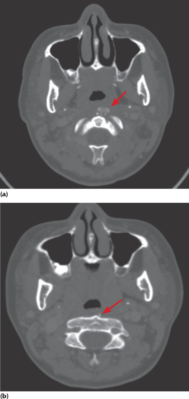
(a). CT scan of neck (axial view) showing focal prevertebral soft tissue thickening at C1 and C2 levels measuring one cm in full thickness. Arrow shows soft tissue calcification anterior to C1 and C2 levels. (b): CT scan of neck (axial view) shows reduced soft tissue thickening at C1 and C2 levels measuring 0.5 cm.