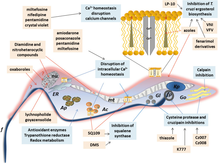
Figure 4.
Schematic representation of a trypomastigote of Trypanosome cruzi and the main cellular targets of the investigational compounds in the pre-clinical phase of development.
Notes: The selected targets are in blue boxes and compounds are in rose boxes. Organelles: Kp: kinetoplast, f: flagellum, fp: flagellar pocket, mt: mitochondrion, nu: nucleus, ER: endoplasmic reticulum, Go: golgi apparatus, Ac: acidocalcisomes, Gl: glycosomes, Ap: autophagosomes. The drawing is based on data from Benaim et al and Vannier-Santos et al.163,228

