
Fig. 5.
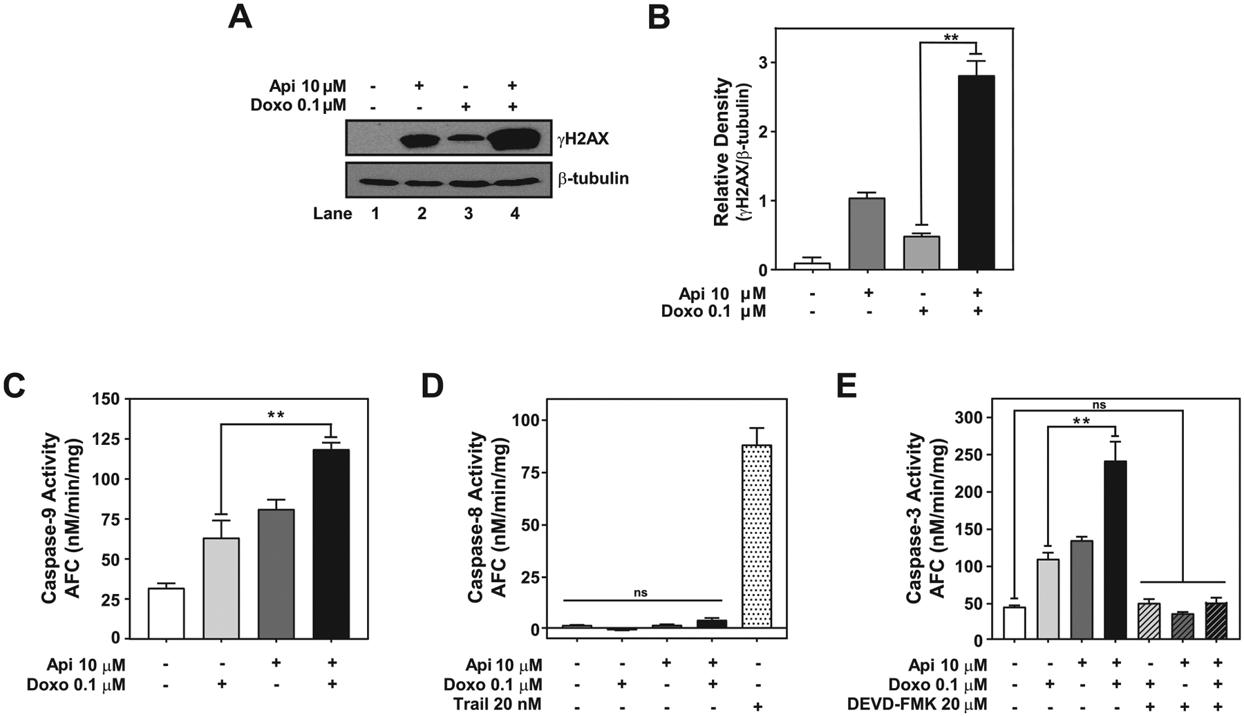
Apigenin enhances doxorubicin cytotoxicity by inducing DNA damage and activation of the intrinsic apoptotic pathway. TNBC MDA-MB-231 spheroids treated with DMSO, 0.1 μM doxorubicin, 10 μM apigenin or simultaneously with doxorubicin and apigenin for 6 days were used to obtain protein lysates. (A) An equal amount of protein lysates from spheroids were analyzed by western blotting using anti-γH2AX antibodies. The same membranes were re-blotted with anti-β-tubulin antibodies, used as the loading control. (B) Relative γH2AX to β-tubulin density of data shown in (A). (C) Protein lysates from spheroids were used to evaluate caspase-9 activity by LEHD-AFC assays. (D) Protein lysates from spheroids were used to evaluate caspase-8 activity by IETD-AFC assays. Protein lysate from spheroids treated with 20 nM TRAIL for 6 days were used as a positive control to evaluate the activation of caspase-8. (E) Caspase-3 activity was evaluated by DEVD-AFC assays in protein lysates from spheroids pretreated for 2 h with DMSO or 20 μM DEVD-FMK prior to treatment with 0.1 μM doxorubicin, 10 μM apigenin or simultaneously with doxorubicin and apigenin for 6 days. Data represent mean ± SEM, N = 3. **p < 0.001, compared to single treatment with doxorubicin, ns indicates no statistical difference.