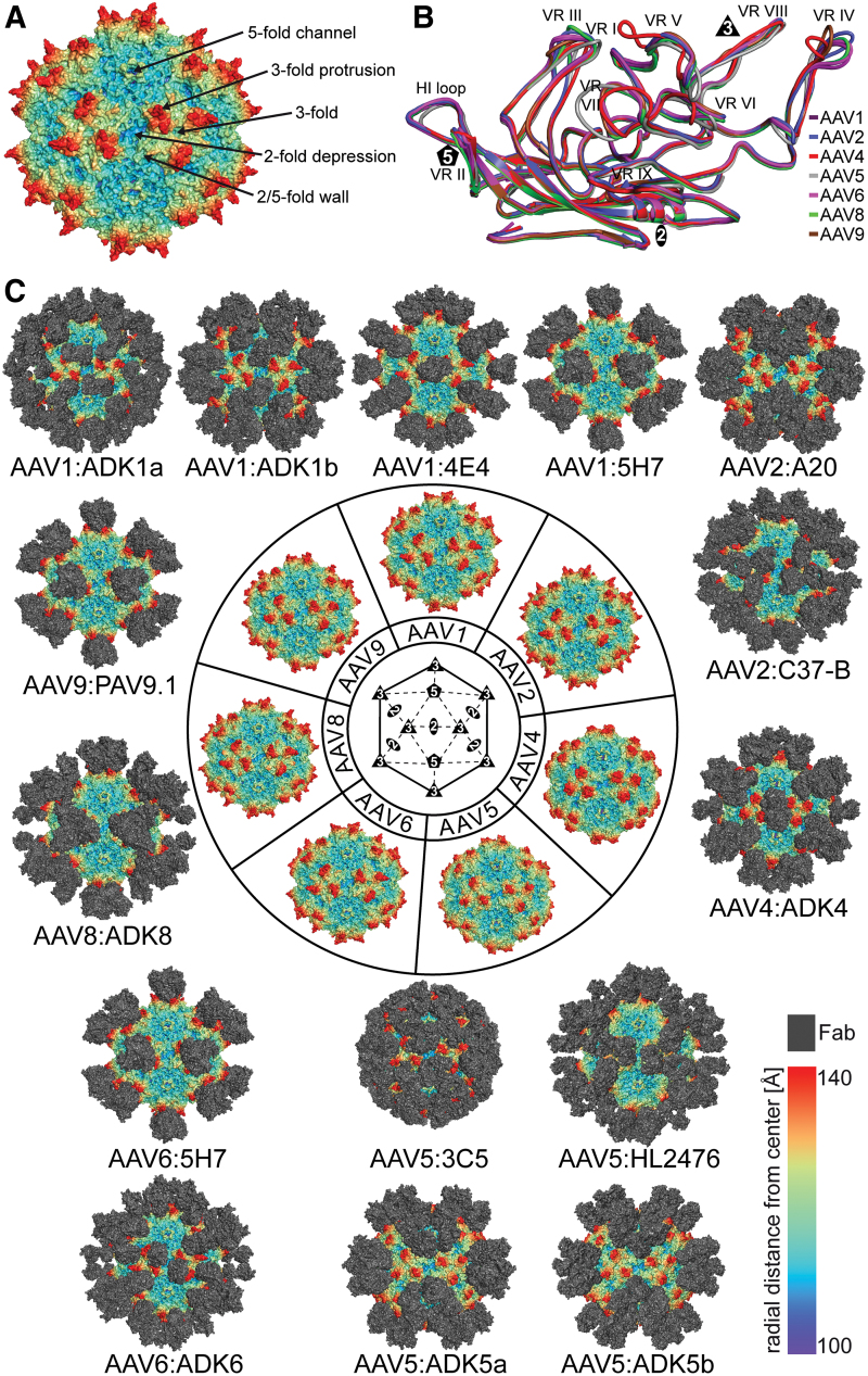
FIG. 1.
Antigenic Interactions of Dependoparvoviruses. (A) Capsid surface representation of AAV2. The approximate locations of the 2-, 3-, and 5-fold axes are indicated along with the 2/5-fold wall and 3-fold protrusions. (B) Structural superposition of VP monomers from different AAV serotypes. The VRs are labeled. (C) Depiction of the cryo-reconstructed density maps of the AAV capsids complexed to the Fab portion of monoclonal antibodies. The serotypes shown are AAV1, 2, 4, 5, 6, 8, and 9, located at the center of the figure. Generic or specific Fab models are docked onto the viral capsids at the appropriate locations identified by the cryo-reconstructed maps. These epitopes span the 2-fold, 3-fold, and 5-fold regions together with the 2/5-fold wall. In (A) and (C) the viral capsids are colored radially, such that regions closest to the center are colored purple, and those farthest away are colored red, and the Fabs are colored gray, as indicated by the scale bar (bottom right). These images were generated using PyMol (31). AAV, adeno-associated virus; Fab, fragment antigen binding; VP, viral protein; VR, variable region. Color images are available online.

