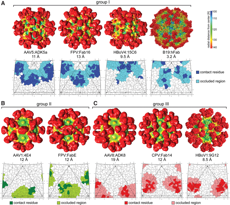
FIG. 5.
Commonalities in Parvovirus Antigenic Epitopes. Example cryo-EM density maps and 2D Roadmaps are shown, viewed down the 2-fold axis of symmetry, for common Fab binding regions for the dependoparvoviruses, protoparvoviruses, bocaparvoviruses, and erythroparvoviruses. (A) Group I—antibodies binding at or around the 5-fold symmetry axis and the 2/5-fold wall. (B) Group II—antibodies binding on the 2-fold facing wall of the 3-fold protrusions across the 2-fold axis. (C) Group III—antibodies binding at or near the 3-fold protrusions. The density maps are colored radially such that regions closest to the center of the capsid are colored blue, and those farthest away are colored red. Capsid density is shown mostly in blue, yellow, and green, while that corresponding to the Fabs are in red (see color key at the top right hand corner). The map images were generated using UCSF-chimera (95). The roadmap projections were generated with Radial Interpretation of Viral Electron density Maps (133). The viral asymmetric unit, with the 5-fold axis (pentagon), the 2-fold axis (ellipse), and the two 3-fold axes (triangles), is shown. Contact and occluded residues are indicated by different color shades. 2D, two-dimensional; cryo-EM, cryo-electron microscopy; RIVEM, Radial Interpretation of Viral Electron density Maps. Color images are available online.

