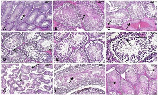
Figure 8.
H&E-stained testicular sections. Effect of exposure to Ag-NPs and/or Zn-NPs on the microscopic appearance of testicular tissue. (A) Testis of control rat showing normal seminiferous tubules with densely packed spermatogonial cells’ layers, spermatogenesis and normal sperms in the lumen (arrow). (B) Testis of Zn-NPs administered rat showing normal seminiferous tubules with active spermatogenesis. (C–G) testis of Ag-NPs administrated rat showing; (C) Thickening of the testicular capsule (arrow) and congested capsular blood vessels with thickening and edema (dotted arrow) of their walls, (D) Disorganized spermatogonial cells’ layers and detached germinal epithelium (dotted arrow) from the basement membrane of the seminiferous tubules and mild interstitial edema (arrow), (E) Degeneration, necrosis and nuclear pyknosis of the spermatogonial cells, (F) Defective spermatogenesis and presence of multinucleated spermatid giant cells (arrow), (G) Seminiferous tubules appeared with irregular contour (arrow) of seminiferous tubules with marked wide spaces between them. (H and I) Testis of Ag-NPs and Zn-NPs co-administered rat showing near to normal appearance of seminiferous tubules with active spermatogenesis, active sperms (dotted arrow) in the lumen of seminiferous tubules and spermatogonial cells (arrow).

