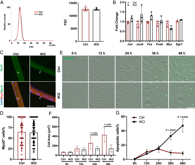
Fig. 3.
Loss of Paxbp1 impaired the cell growth of MuSCs without affecting the initial early activation. (A) Representative forward scatter (FSC) distribution of FISCs from a Ctrl and an iKO mouse (Left) and quantification of the mean FSC for two types of FISCs (Right) (n = 5 mice per group). (B) Measurement of relative mRNA expression of selected IEGs by qRT-PCR using FISCs from Ctrl and iKO mice (n = 3 mice per group). (C) Freshly isolated myofibers from Ctrl and iKO mice were cultured for 8 h before fixation followed by immunostaining for MyoD and Pax7. White arrows indicate ASCs on myofibers. (D) Quantification of the percentage of MyoD+ ASCs over Pax7+ ASCs in C; ∼90 fibers from n = 3 mice per group were used for quantification. (E) Live-cell imaging was conducted on FISCs from control and iKO mice grown in culture together with CellEvent Caspase-3/7 Green Detection Reagent for 72 h. Representative snapshots at the indicated time points are presented. (F) Quantification of the mean cell size in E by Fiji (ImageJ): At each time point, >100 cells from n = 3 mice per group were measured. (G) Quantification of the percentage of Caspase3/7-positive cells in E (n = 3 mice per group). Data are presented as mean ± SD. (Scale bars, 50 µm.)

