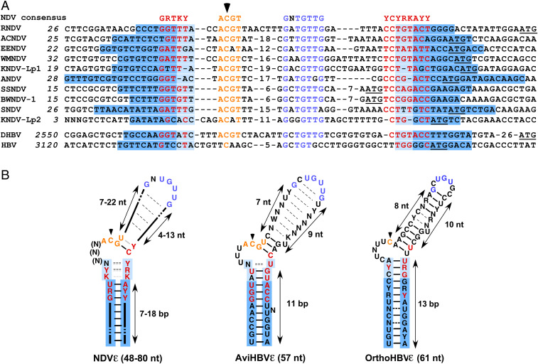
Fig. 2.
Comparison of nackednaviral and hepadnaviral ε elements. (A) Alignment of nackednaviral and hepadnaviral ε sequences. Colored letters indicate conserved sequence elements in lower stem (red), initiation region (orange), and apical region (blue). Base-paired lower stem regions are shaded in blue, and the variably base-paired tip of the lower stem is in light blue. A black arrowhead denotes the intiation site. The smORF1 start codons of nackednaviruses and the core start codons of HBV and DHBV are underlined. Internal numbers denote nt not depicted in the figure. Italic numbers refer to genomic nt positions (2). African cichlid nackednavirus (ACNDV), European eel nackednavirus (EENDV), Western mosquitofish nackednavirus (WMNDV), Lucania parva killifish nackednavirus-1 and -2 (KNDV-Lp1, KNDV-Lp2), Astatotilapia nackednavirus (ANDV), sockeye salmon nackednavirus (SSNDV), baby whale nackednavirus-1 (BWNDV-1), and stickleback nackednavirus (SNDV). (B) Consensus ε structures of nackednaviruses (Left), avihepadnaviruses (Middle), and orthohepadnaviruses (Right). Dashed lines between nt indicate nonconserved, facultative bp, and gray lines indicate potential noncanonical bp.

