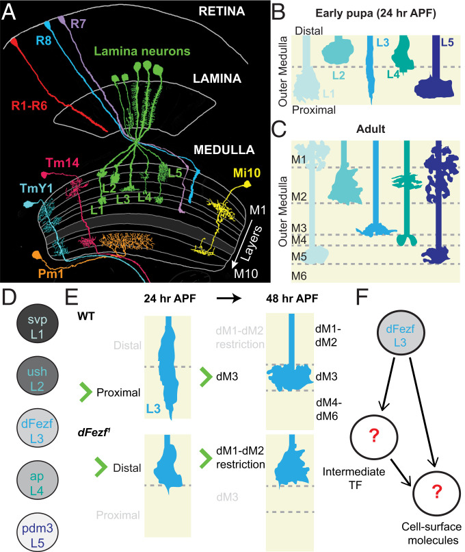
Fig. 1.
The D. melanogaster visual system and lamina neuron layer specificity. Schematics illustrating the various morphologies and layer-specific innervation patterns found throughout the fruit fly optic lobe, as well as a synopsis of the findings from Peng et al. (12). (A) Visual information is received by the retina and relayed to four downstream neuropil known as the lamina, medulla, lobula, and lobula plate (lobula and lobula plate are not shown in the diagram). The medulla neuropil consists of 10 layers (M1 to M10). The M1 to M6 layers constitute the outer medulla, while the M8 to M10 layers constitute the inner medulla, both of which are separated by the serpentine layer (M7). More than 100 morphologically distinct neurons innervate specific layers throughout the medulla, wherein they form stereotyped connections. Reprinted by permission from ref. 28, Springer Nature: Cell and Tissue Research, copyright (1989). (B and C) The L1 to L5 lamina neurons are major input neurons into the medulla that establish distinct innervation patterns throughout layers M1 to M5 of the outer medulla. (B) During early pupal development (24 h APF), L1 to L5 growth cones innervate the primordial outer medulla, which is partitioned into the distal and proximal broad domains at this stage. L1 to L5 establish primitive, overlapping targeting patterns before arborizing in unique patterns illustrated in C. (D) Each lamina neuron expresses a unique transcription factor, which we hypothesize confers the layer specificity for each cell type. (E) Summary of findings in Peng et al. (12). Growth cones of dFezf1 mutant L3 neurons are initially restricted to the distal domain and later terminate in superficial layers (dM1 to dM6 are the developing layers M1 to M6 observed in the adult [C]). We hypothesize dFezf regulates the expression of cell surface genes either directly or via intermediate regulators (F).

