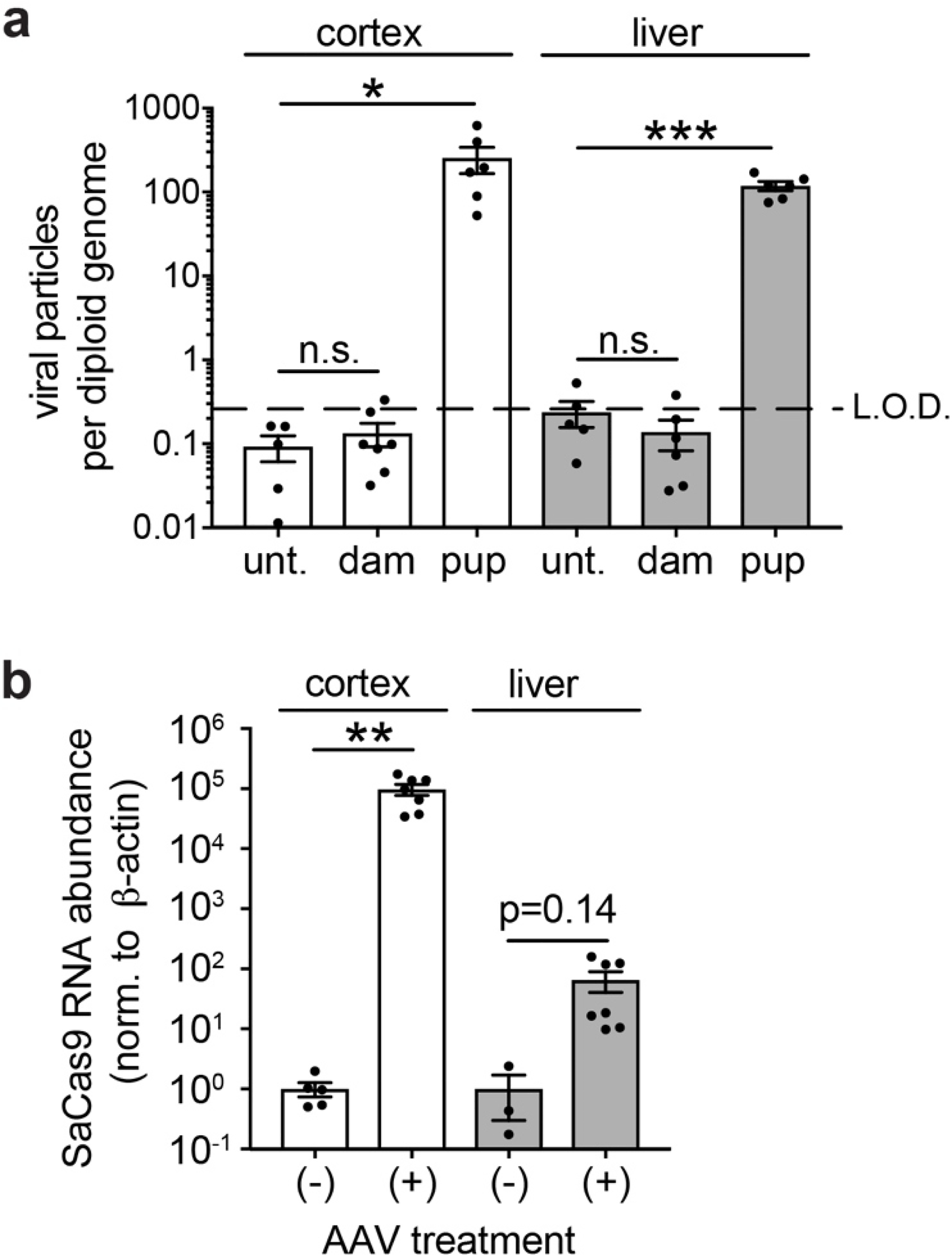
Extended Data Figure 7. Evaluation of viral transduction in tissues from injected mice and their dams.

a. qPCR quantification of viral DNA (SaCas9 TaqMan probes) in cortex and liver of age matched untreated animals, P60 dams whose pups were injected i.c.v. at E15.5, and P60 mice that were dual injected i.c.v. at E15.5+P1. Data normalized to Eif4a2 (representing a gene with 2 copies per diploid genome). Limit of detection (LOD) determined by performing serial dilutions with known quantities of AAV particles spiked into gDNA samples from untreated mice. * P<0.05, ** P<0.01, *** P<0.001.
b. Expression of SaCas9 mRNA in cortex and liver from same animals as (a). ΔΔCt method, normalized to ß-actin.
