Figure 1.
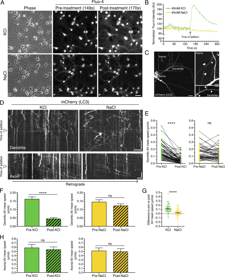
Neuronal depolarization dampens AV motility specifically in dendrites and not in axons. (A) Rat hippocampal neurons (13 DIV) were loaded with the calcium indicator Fluo-4 and treated with KCl to depolarize the neurons or with NaCl as an osmotic control. Scale bar, 50 µm. (B) Quantitation of mean Fluo-4 fluorescence intensity before and after KCl or NaCl addition. (C) Distribution of mCherry-LC3–positive AVs in dendrites and axons of primary rat hippocampal neurons (13 DIV). Image with soma; scale bar, 10 µm. Magnified images of axons and dendrites with arrowheads denoting mCherry-LC3–positive puncta; scale bars, 5 µm. (D) Kymographs of LC3 motility along a dendrite or an axon of a rat hippocampal neuron. Retrograde direction is from left to right. Arrowheads denote time of KCl or NaCl addition. Horizontal bars, 5 µm. Vertical bars, 1 min. (E–G) Quantitation of AV mean speed in dendrites before and after KCl or NaCl addition (E, paired points represent a single AV before and after salt addition; F, mean ± SEM; G, difference in AV mean speed before and after salt addition; KCl, n = 68 AVs from 20 neurons from three independent experiments, 13–14 DIV; NaCl, n = 55 AVs from 20 neurons from three independent experiments, 13–14 DIV; E and F, paired t test; G, unpaired t test; ****, P ≤ 0.0001). (H) Quantitation of AV mean speed in axons before and after KCl or NaCl treatment (mean ± SEM; n = 56–125 AVs from 8–10 neurons from three independent experiments; 13–14 DIV; unpaired t test).

