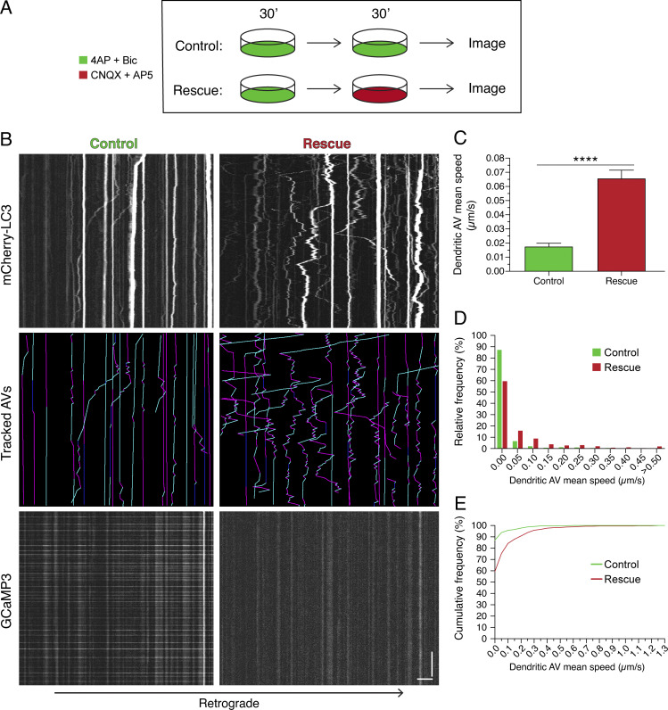
Figure 4.
Activity-dependent dampening of AV motility in dendrites is reversed by silencing neurons. (A) Schematic of experimental paradigm. (B) Kymographs of mCherry-LC3, tracked AVs (magenta denotes retrograde, cyan denotes anterograde, and dark blue denotes stationary segments), and GCaMP3 from the same dendrite after the treatments described in A. Horizontal bar, 5 µm. Vertical bar, 1 min. (C–E) Quantitation of AV mean speed in rat hippocampal neurons treated with the experimental paradigm in A (C, mean ± SEM; D, histogram of dendritic AV mean speed; E, cumulative frequency of dendritic AV mean speed; n = 531–641 AVs from 25–26 neurons from three independent experiments; 13–14 DIV; unpaired t test; ****, P ≤ 0.0001).

