Figure 5.
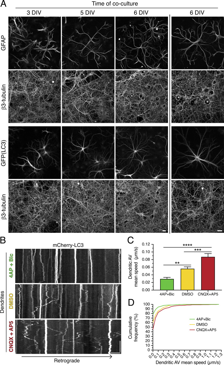
Synaptic activity controls AV dynamics in dendrites of neurons cocultured with astrocytes. (A) Maximum projections of Z-stacks of rat hippocampal neurons (immunostained for neuron-specific β3-tubulin) cocultured with either wild-type mouse cortical astrocytes [immunostained for astrocyte-specific glial fibrillary acidic protein (GFAP)] or GFP-LC3–transgenic mouse cortical astrocytes (immunostained for GFP). Images to the left of the vertical line were taken with a 40× objective, and images to the right of the vertical line were taken with a 63× objective. Asterisks denote the location of neuronal cell bodies within the neurite meshwork. Scale bars, 20 µm. (B) Kymographs of mCherry-LC3 in dendrites of rat cortical neurons cocultured with mouse cortical astrocytes for 7–8 DIV (neurons are a total of 14–16 DIV) and treated for 30 min in 4-AP + Bic, DMSO, or CNQX + AP5. Retrograde direction is from left to right. Horizontal bar, 5 µm. Vertical bar, 1 min. (C and D) Quantitation of AV mean speed in dendrites of cortical neurons cocultured with cortical astrocytes for 7–8 DIV and treated for 30 min in 4-AP + Bic, DMSO, or CNQX + AP5 (C, mean ± SEM; D, cumulative frequency of dendritic AV mean speed; n = 323–368 AVs from 9–10 neurons from two independent experiments; neurons are 14–16 DIV; one-way ANOVA with Tukey’s post hoc test; **, P ≤ 0.01; ***, P ≤ 0.001; ****, P ≤ 0.0001).

