Figure 6.
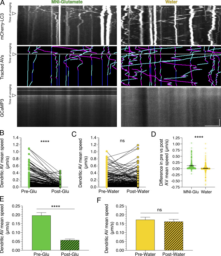
Local stimulation of synaptic activity by uncaging MNI-glutamate (MNI-Glu) reduces AV motility in dendrites. (A) Kymographs of mCherry-LC3, tracked AVs (magenta denotes retrograde, cyan denotes anterograde, and dark blue denotes stationary segments), and GCaMP3 from the same dendrite before and after glutamate (Pre-Glu and Post-Glu, respectively) uncaging (arrowheads denote time of uncaging; water serves as a control). Horizontal bar, 5 µm. Vertical bar, 30 s. (B–F) Quantitation of AV mean speed in dendrites before and after uncaging. (B and C, paired points represent a single AV before and during uncaging; D, difference in AV mean speed before and after uncaging; MNI-glutamate, n = 226 AVs from 22 hippocampal neurons; water, n = 268 AVs from 25 hippocampal neurons; three independent experiments; 13–14 DIV; B and C, paired t test; D, unpaired t test; ****, P ≤ 0.0001). (E and F) Mean ± SEM of all AVs tracked, including those from paired analysis, before and after uncaging (E, n = 272 AVs in pre-uncaging and 277 AVs in post-uncaging from 22 neurons from three independent experiments; 13–14 DIV; F, n = 321 AVs in pretreatment and 346 AVs in post-treatment from 25 neurons from three independent experiments; 13–14 DIV; unpaired t test; ****, P ≤ 0.0001).

