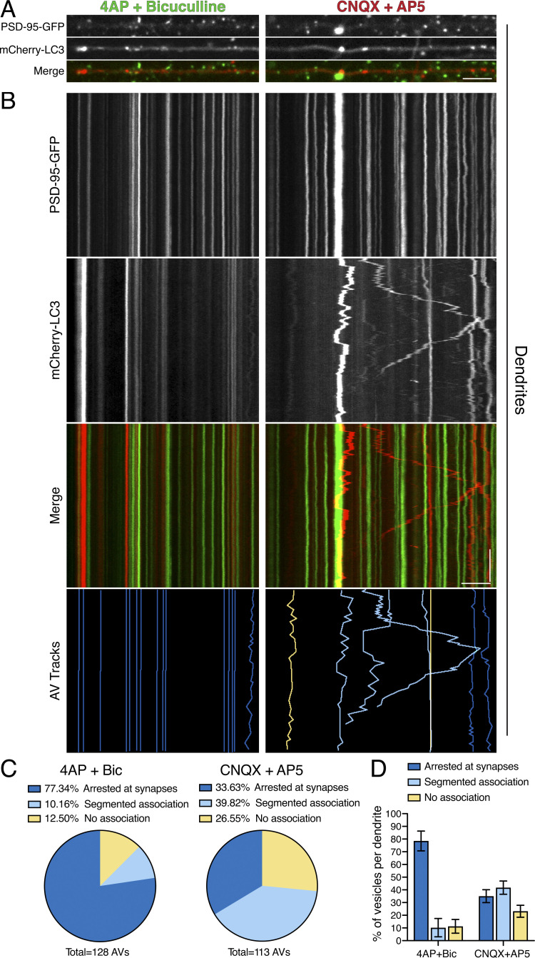
Figure 7.
Synaptic activity arrests AVs at or proximal to post-synaptic compartments. (A and B) En face images (A) and kymograph analysis (B) of PSD-95–GFP, mCherry-LC3, and tracked AVs (dark blue denotes AVs arrested at synapses, light blue denotes AVs with segmented association with synapses, and yellow denotes AVs with no association with synapses) in dendrites of hippocampal neurons. Horizontal bars in A and B, 5 µm. Vertical bar in B, 1 min. (C) Quantitation of individual AV association with synapses as a percentage of total AVs tracked (4-AP + Bic, n = 128 AVs from 10 neurons; CNQX + AP5, n = 113 AVs from 11 neurons; two independent experiments; 13–16 DIV). (D) Quantitation of percentage of AV association with synapses on a per dendrite basis (mean ± SEM; n = 10–11 neurons from two independent experiments; 13–16 DIV).

