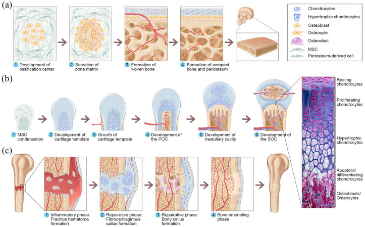
Figure 1.
Overview of IMO and ECO during embryonic bone development and fracture healing: (a) IMO follows four steps. Step 1: MSCs undergo condensation and form ossification centers. MSCs within the areas of condensation lead to the development of capillaries and osteoblasts. Step 2: Osteoblasts secrete osteoid, which then entraps the osteoblasts, and the osteoblasts transform into osteocytes. Step 3: Osteoid secreted around the capillaries result in trabecular matrix formation, while osteoblasts on the surface of the spongy bone become the periosteum. Step 4: The periosteum then creates compact bone superficial to the trabecular bone. The trabecular bone crowds blood vessels, which eventually condense into red marrow. (b) ECO follows six steps during embryonic bone development. Step 1: MSC condensation. Step 2: MSCs within the areas of condensation differentiate into chondrocytes to form the cartilage template of the future long bone, and MSCs in the periphery form the perichondrium. Step 3: Chondrocytes in the center of the template undergo hypertrophy, while cells in the periphery undergo direct osteogenic differentiation to form a periosteal collar of compact bone around the cartilage template. Step 4: Hypertrophic chondrocytes secrete osteogenic and angiogenic factors that initiate cartilage matrix mineralization and blood vessel invasion, resulting in POC formation. Step 5: The diaphysis elongates, and a medullary cavity forms as ossification continues. Step 6: After this initial bone formation, the same sequence of events occurs in the epiphyseal regions, leading to SOC formation, and (c) The healing of fractures follows three consecutive and overlapping phases. Inflammatory phase: Approximately 6–8 h after the fracture, a hematoma is formed at the fracture site. Reparative phase: Within approximately 48 h after the fracture, chondrocytes from the periosteum and marrow create an internal callus between the two ends of the broken bone and an external callus around the outside of the break. MSCs from the periosteum directly differentiate into osteoblasts, thereby stimulating appositional bone growth and enveloping the defect. Over the next several weeks, the cartilage in the calli is replaced by woven bone via ECO. Remodeling phase: The woven bone remodels into lamellar bone through osteoclast-osteoblast coupling, and the healing process is complete. The histological image of the epiphyseal plate of a growing long bone was adapted from Human Anatomy, sixth edition (Copyright © 2011 Pearson Education, Inc., Figure 6.12).

