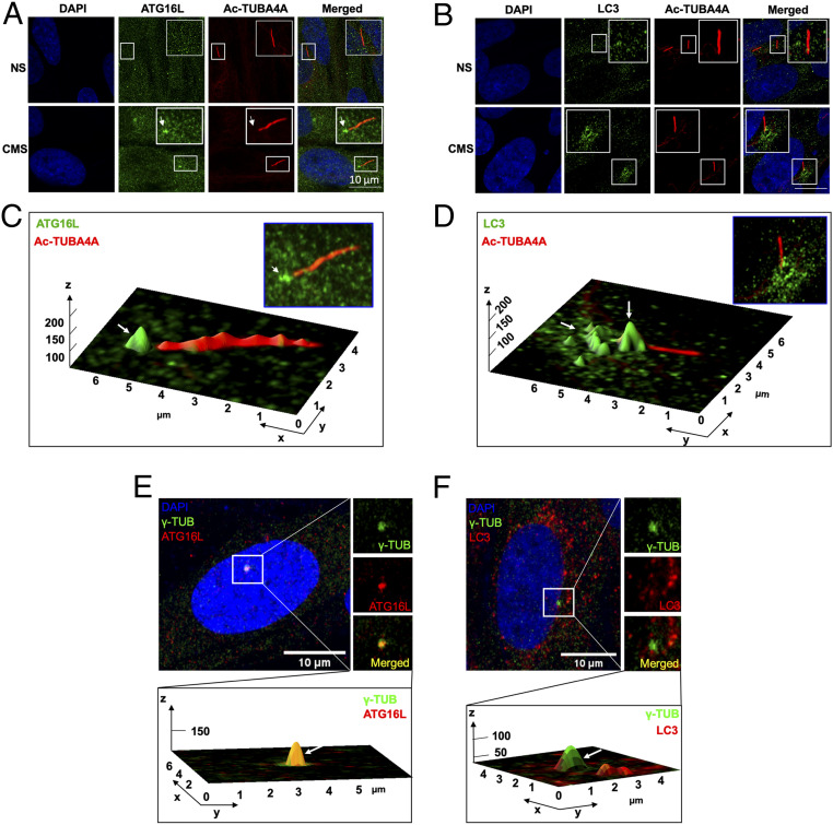
Fig. 2.
ATG16L and LC3 localize at the basal body of the PC upon CMS. (A and B) Representative immunostaining of ATG16L (A, green) and LC3 (B, green) in TM cells exposed for 24 h to CMS (8% elongation). PC were stained using acetylated TUBA4A (red); DAPI was used to stain nuclei. (C and D) Interactive three-dimensional (3D) surface plot analysis visualizing the accumulation of ATG16L (C, green) and LC3 (D, green) in the basal body of PC of the CMS cells (white arrows). (E and F) Representative immunostaining of ATG16L (A, red) and LC3 (B, red) with ɣ-tubulin (green), a maker for the basal body of PC, in TM cells exposed for 24 h to CMS (8% elongation). (Bottom) Interactive 3D surface plot analysis visualizing the colocalization of ATG16L or LC3 with ɣ-tubulin (white arrows).

