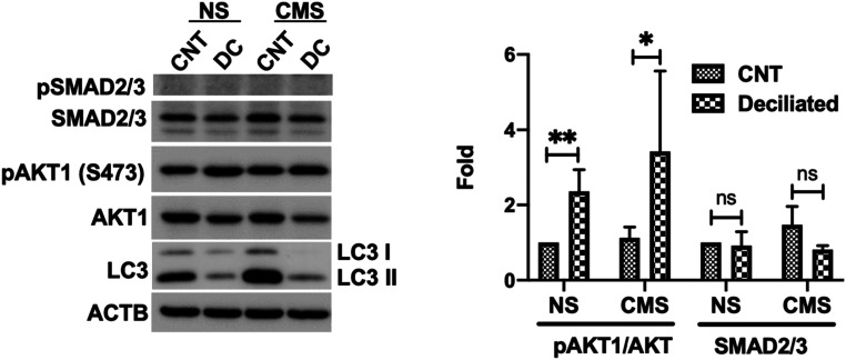
Fig. 6.
AKT phosphorylation is increased in deciliated cells. Cells were treated in 4 mM CH for 3 d and subjected to CMS after CH removal (8% elongation, 24 h). Protein levels of AKT1, pAKT1, SMAD2/3, and LC3 were evaluated by WB. Data are shown as the mean ± SD (n = 3). **P < 0.01; *P < 0.05 (two-tailed unpaired Student’s t test); ns, not significant.

