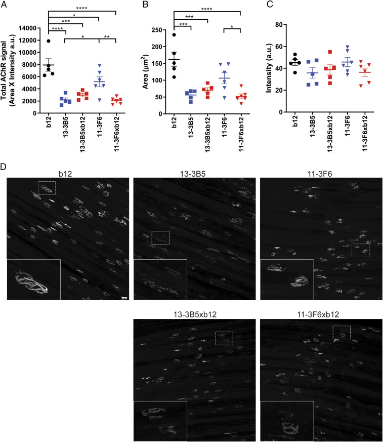
Fig. 3.
Monovalent and bivalent MuSK antibodies impair NMJ morphology to a different extent. To visualize the postsynaptic NMJ, AChRs were stained with AF488-BTX on diaphragm muscle preparations. Twenty randomly selected NMJs per diaphragm were analyzed and averaged. (A) Total AChR signal was calculated by multiplying the positive area with the average intensity per NMJ. Both bivalent and monovalent MuSK antibodies significantly reduced the total AChR signal compared with the b12-treated mice. Bivalent 13–3B5 and monovalent 11–3F6xb12 reduced the total AChR signal to a greater extent compared with monospecific 11–3F6. Bivalent 13–3B5 did not significantly differ from monovalent 13–3B5xb12. (B) To assess the size of the NMJs, a threshold was applied, and the area of the positive signal was quantified. Exposure to monovalent MuSK antibodies or bivalent 13–3B5 resulted in less signal reaching the threshold and therefore smaller NMJs. Monovalent 11–3F6xb12 also caused significantly smaller NMJs compared with bivalent 11–3F6. (C) The average intensity of AChR staining surpassing the threshold did not significantly differ between groups. (D) Representative maximum projections per condition with insets. (Scale bar, 25 µm.) 11–3F6 and 11–3F6xb12 n = 6, 13–3B5, 13–3B5xb12 and b12 n = 5. Data represent mean ± SEM. One-way ANOVA with Šidák-corrected comparisons for all parameters: *P <0.05, **P <0.01, ***P <0.001, ****P <0.0001.

