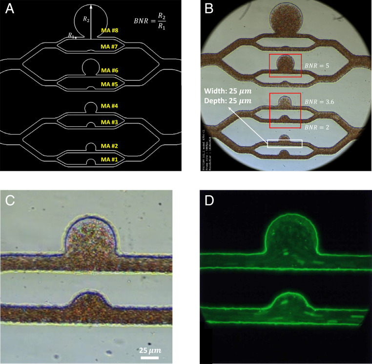
Fig. 2.
Design and microscopic images of the microfluidic platform, MAOAC. (A) Schematic diagram of the MA PDMS channels with different sizes. The size of the MA is characterized by the BNR, which is defined as the largest dimension of the MA body () divided by the diameter of feeding vessel (). The BNR of MA on MAOAC is varied from 1.5 to 12. The cross-section of the microchannel at the inlet and outlet portions is a square with an edge size of 25 . (B) An overview of a bright-field image illustrating blood flow in the microchip. (More details can be seen in Movie S1.) (C) A higher-magnification view of the bright-field image of two MA channels (BNR = 2 and BNR = 3.6). (D) A fluorescence-stained image of the same two MA channels shown in C. (More details can be seen in Movie S2.) AIV is used to extract velocity and pressure fields from the bright-field video images focusing on MAs with three typical sizes, namely, BNR = 2, 3.6, and 5, which are highlighted by the red rectangles in B. For MAs with BNR = 2 and 3.6, we perform platelet tracking on the fluorescence-stained video (D) for validation of AIV.

