


Figure 2. Microscopic analysis of efferocytosis.
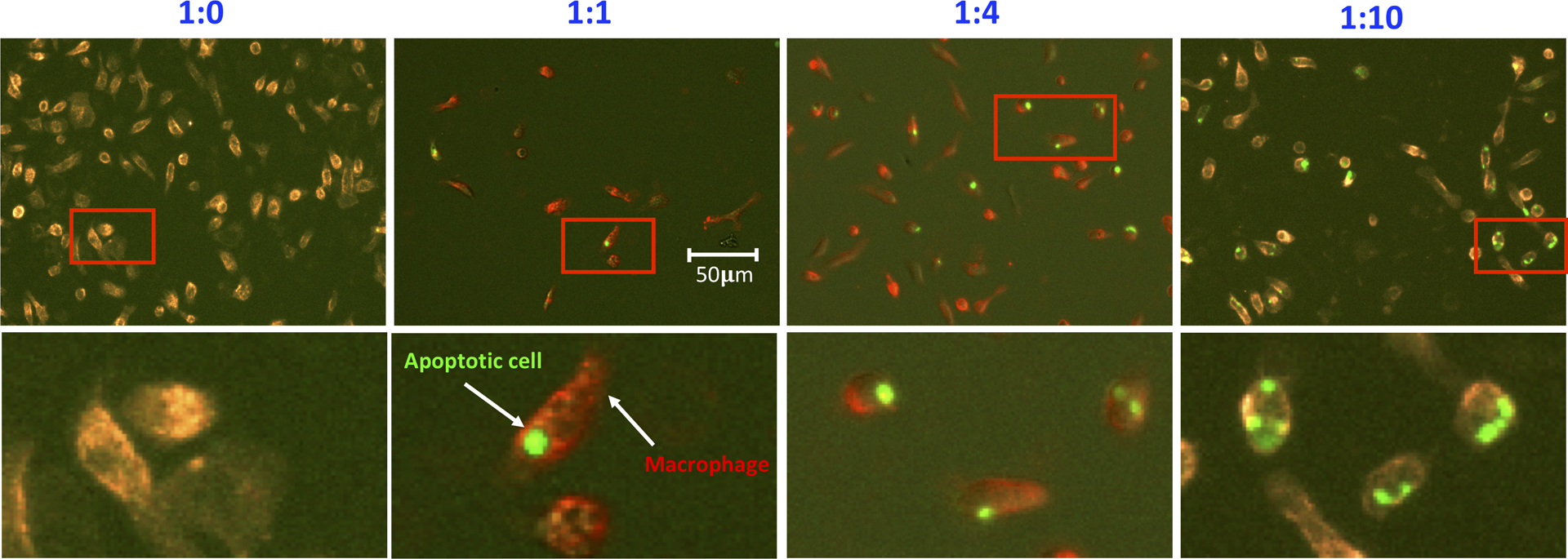
Efferocytosis was prepared as in Figure 1. Phagocytic macrophages were stained in situ on plate with CD11b-PE, fixed with paraformaldehyde, and evaluated. Images were acquired using the fluorescent microscope and analyzed with the software associated with the microscope. Representative insertions were digitally enlarged and shown below in each image.