Figure 4.
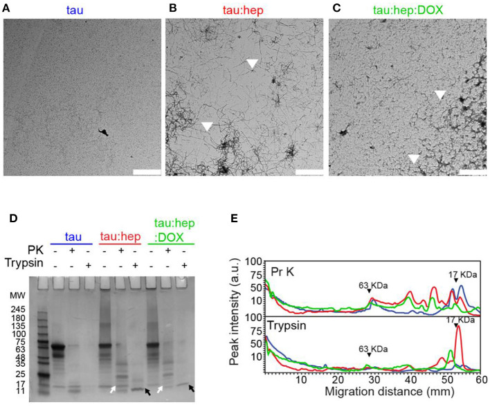
Doxycycline interferes with heparin-induced tau fibril formation. (A–C) Transmission electron microscopy (TEM) of different 2N4R tau samples incubated at 37°C under orbital agitation and harvested after 168 h of incubation. Scale bar corresponds to 2 μm. (D) Partial digestion profile of tau samples incubated in the same conditions as A, treated and not treated with 1 μg/ml proteinase K and with 0.0125% trypsin. Digestion products were resolved in a 12% tris-glycine gel stained with colloidal Coomasie Blue. Molecular weight marker in kDa. Comparison between digestion products of tau: hep and tau:hep:DOX aggregates obtained by PK (white arrows) and Trypsin (black arrows) proteolysis. SDS-PAGE gel image was carefully selected (from at least three experiments) to be representative. (E) Densitometric analysis of the SDS-PAGE gel B was performed by using Image J 1.47v software.

