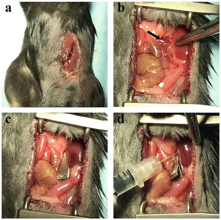
Fig. 1.
Different steps of the retrograde renal vein injection. (a) Incision in the upper-left quadrant of the mouse abdomen. (b) Exposure of the kidney and identification of the renal vein (arrow). (c) Positioning of the clamp with the kidney appearing darker after a proper clamping. (d) Injection of the rAAV solution in the segment of the vein between the clamp and the kidney

