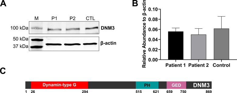
Fig. 7.
A. Expression of DNM3 protein in patient and control fibroblasts. Image shows a representative Western blot image to detect the presence of DNM3 or β-actin protein in lysates extracted from Patient 1 (P1), Patient 2 (P2) or control (CTL) fibroblasts. Molecular weight protein markers (M) are shown in the left lane. B. Quantification of DNM3 expression relative to β-actin from three independent experiments. C. Protein domains of DNM3 (UniProt# Q9UQ16). “Dynamin-type G”: Dynamin-type guanine nucleotide-binding (G) domain, “PH”: Pleckstrin homology domain, “GED”: GTPase effector domain [9].

