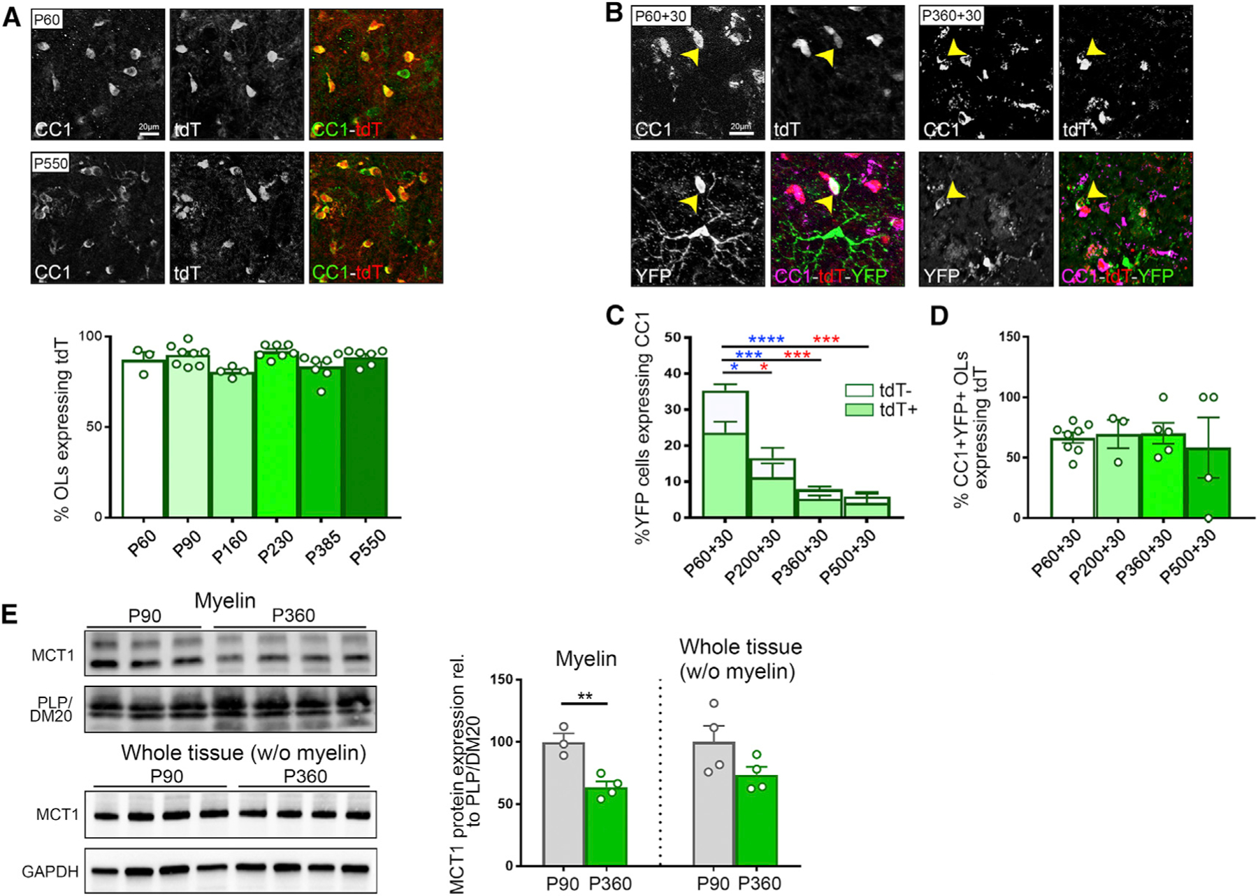
Figure 1. Loss of Oligodendrocyte (OL) MCT1 Protein Expression with Aging.

(A) Immunostaining and quantification of CC1+ OLs expressing MCT1-tdTomato (tdT) between the ages of P60 and P550 (n = 3–8). Data are represented as mean ± SEM.
(B) Evaluation of the ability of newly generated OLs to express tdT. P60-, P200-, P360-, and P550-old Pdgfrα-CreER-RosaYFP-MCT1td-Tomato mice were injected with tamoxifen to induce YFP labeling of OPCs and were euthanized 30 days later. Immunostaining highlights differentiated CC1+ OLs expressing tdT and YFP at the ages of P60+30 and P360+30. Images are representative of n = 5–8.
(C and D) Quantification of the fraction of newly generated OLs (# CC1+YFP+/# YFP+, full bars, statistics in blue) and the fraction of newly generated OLs expressing tdT (# CC1+YFP+MCT1tdTomato+/# YFP+, light green bars, statistics in red) reveals a significant decrease with aging (C) (n = 4–8, *p < 0.05, ***p < 0.001, ****p < 0.0001, one-way ANOVA with Tukey’s multiple comparison test). The fraction of newly differentiated OLs that turn on tdT expression (# CC1+YFP+MCT1tdTomato+/# CC1+YFP+) is unaffected by aging (D) (n = 3–8). Data are represented as mean ± SEM.
(E) Western blot analysis of MCT1 protein in purified myelin fractions reveals a 35% reduction at P360 compared with P90 (n = 3–4, **p < 0.01, Student’s t test), whereas MCT1 protein expression in the ‘‘whole tissue (without [w/o] myelin)’’ fraction is unchanged. Data are represented as mean ± SEM.
