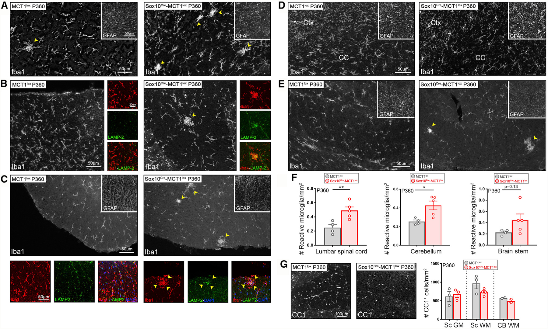
Figure 5. Increased Microglial Reactivity in the CNS WM of Aged Sox10Cre-MCT1lox Mice.

(A–E) Microglial Iba1+ reactivity is enhanced in the cerebellum (A), DCST (B), and ventral spinal cord WM (C) of P360 Sox10Cre-MCT1lox mice compared with MCT1lox controls (arrowheads). Iba1+ immunoreactivity co-localized with LAMP-2 (Mac 3), confirming that microglia acquire a more reactive phenotype (small panels taken from larger images in B and C). There was no change in GFAP immunoreactivity (insets). Microglial Iba1+ and astrocyte GFAP+ reactivity in the Ctx and CC of P360 Sox10Cre-MCT1lox is unaffected compared with MCT1lox controls (D). Images are representative of n = 4–5.
(F) Quantification of changes in microglial Iba1+ reactivity reveals a 2-fold increase in the spinal cord and a 1.7-fold increase in the cerebellum of P360 Sox10Cre-MCT1lox mice compared with MCT1lox controls (spinal cord: **p < 0.01; cerebellum: *p < 0.05; n = 4–5, Student’s t test). No change in the number of reactive microglia was observed in the brain stem (n = 4–5). Data are represented as mean ± SEM.
(G) There are no differences in the number of CC1+ OL cell number in the GM spinal cord (Sc GM), WM spinal cord (Sc WM), and WM cerebellum (CB WM) between P360 Sox10Cre-MCT1lox and MCT1lox mice (n = 2–4). Data are represented as mean ± SEM.
