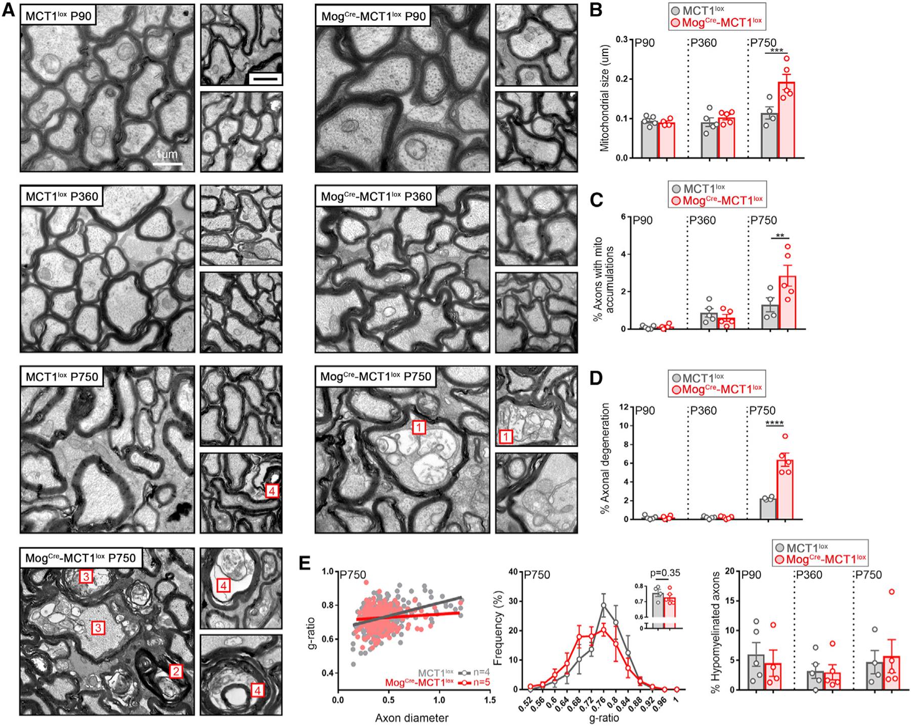
Figure 7. Age-Dependent Axonal Degeneration in Optic Nerves Isolated from MogCre-MCT1lox Mice.

(A) Electron microscopy images from P750 optic nerves from MogCre-MCT1lox reveal the presence of swollen mitochondria (1), accumulation of redundant myelin loops (2), and active degenerating (3) as well as degenerated (4) axons containing multivesicular structures. These morphological changes were only rarely observed in the age-matched MCT1lox controls.
(B–D) At P750, mitochondrial size is 1.7-fold increased in the MogCre-MCT1lox mice compared with the MCT1lox controls (B), the accumulation of intra-axonal mitochondria is 2.2-fold increased in the MogCre-MCT1lox mice compared with the MCT1lox controls (C), and the extent of axonal degeneration is 2.9-fold increased in the MogCre-MCT1lox mice compared with the MCT1lox controls (D) (n = 4–5, **p < 0.01, ***p < 0.01, ****p < 0.0001, two-way ANOVA with Šidák’s multiple comparison test). Data are represented as mean ± SEM.
(E) Evaluation of myelin thickness in P90- and P750-old MogCre-MCT1lox mice does not reveal changes in g-ratio, g-ratio distribution, or percentage of hypomyelinated axons (g-ratio > 0.85 [n = 4–5]). Data are represented as mean ± SEM.
