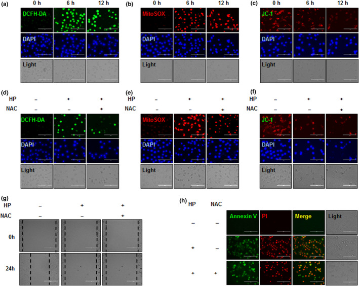
Figure 3.

Effect of hemp peptides on cellular and mitochondrial reactive oxygen species and mitochondrial membrane permeability in Hep3B cells. Hep3B cells were treated with 10 mg/ml hemp peptides for 0, 6, and 12 hr. (a) Cellular reactive oxygen species (ROS) levels were detected by DCFH‐DA (green) and DAPI (blue) staining. (b) Mitochondrial ROS levels were measured by fluorescence microscopy using MitoSOX (red) and DAPI (blue) staining. (c) Mitochondrial membrane potential (MMP) was detected by JC‐1 assay. Hep3B cells were pretreatment with 5 mM NAC for 30 min prior to 10 mg/ml hemp peptide treatment for 12 hr. (d) Cellular ROS levels determined by DCFH‐DA (green) and DAPI (blue) staining. (e) ROS levels at the mitochondria were measured by fluorescence microscopy using MitoSOX (red) and DAPI (blue) staining. (f) Mitochondrial membrane potential detected by JC‐1 assay. (g) Cell migration determined by wound healing assay. (H). Apoptosis levels determined by fluorescence microscopy of Annexin V‐FITC (green) and PI (red). Scale bars represent 100 μm for all images
