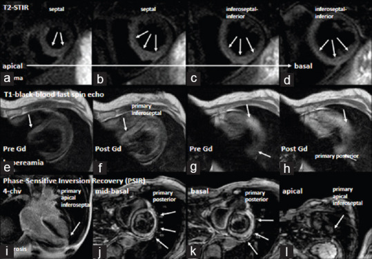
Figure 2.

Documentation of cardiac magnetic resonance imaging findings: Documentation of a predominant edema in the septal and inferior apical to basal left ventricular segments (a-d). Arrows label regions of increased T2 ratio in in T2-STIR (short tau inversion recovery) sequences. Documentation of hyperemia by T1-BBS sequences (black blood fast spin) by early gadolinium enhancement (e-h). Arrows label regions of increased early enhancement (e, g-prior to contrast, f, h-after contrast). Documentation of fibrosis by late gadolinium enhancement in phase sensitive inversion recovery (PSIR) sequences (i-4-chamber view, j-l-short-axis views). Arrows label regions of transmural and subepicardial delayed enhancement
