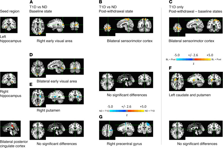
Figure 3. Seed-based functional connectivity maps.
Seed masks were extracted from the FreeSurfer parcellation of the N27 brain template. Three seed regions reached statistical significance (P < 0.01) (left and right hippocampi and bilateral posterior cingulate cortex). The functional connectivity (FC) of these seed regions was higher (yellow) or lower (blue) compared with 6 different brain regions (Table 2). At baseline (left column), there was appreciably higher FC in nondiabetic (ND) participants relative to that in participants with type 1 diabetes (T1D) (A) between the left hippocampus and the right early visual area and (D) between the right hippocampus and bilateral early visual areas. (E) In contrast, there was significantly (P < 0.01) lower FC between the right hippocampus and right putamen in ND participants. There were no significant FC differences with the bilateral posterior cingulate cortex in the baseline state. Following insulin deprivation (middle column), there was substantially higher FC in ND participants relative to that in participants with T1D (B) between the left hippocampus and bilateral sensorimotor cortices but (G) lower FC between both posterior cingulate cortices and the right precentral gyrus. There were no significant FC differences with the right hippocampus in the postwithdrawal state. Finally, assessing changes between the postwithdrawal and baseline states in participants with T1D (right column), (C) there was considerably decreasing FC between the left hippocampus and bilateral sensorimotor cortices and (F) the right hippocampus and the left caudate/putamen following insulin withdrawal, indicating that insulin deprivation adversely affected FC between these regions in participants with T1D. No significant FC differences with the bilateral posterior cingulate cortex were found in participants with T1D between the baseline and postwithdrawal states. Note that the FC clusters for the bilateral sensorimotor cortex identified in B and C are similar but not identical (see Table 2).

