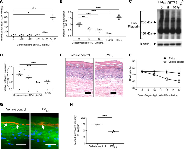
Figure 2. Effects of PM2.5 on FLG and skin barrier function in cultured keratinocytes and organotypic skin.
(A) The percentage of cell death (lactate dehydrogenase release into cell culture media) is increased after exposure to PM2.5. Gene (B) and protein (C and D) expressions of FLG in cultured HEKs were evaluated using reverse transcriptase PCR (RT-PCR) and Western blotting, respectively, and demonstrated reduced FLG mRNA and protein expression in PM2.5-treated cultures. H&E staining (E) and TEWL (F) in organotypic skin. FLG protein expression (G and H) was evaluated in organotypic skin using immunofluorescence staining. Arrows point to FLG staining (shown in red). Wheat germ agglutinin–conjugated FITC (green) was used to stain the cytoskeleton. Nuclei were visualized with DAPI (blue). Data are representative of 3 independent experimental repetitions using 3 different lots of HEKs. The data are shown as the mean ± SEM. n = 3–4 per group. Scale bar: 50 μm. *P < 0.05, **P < 0.01, ***P < 0.001 by 1-way ANOVA with Tukey-Kramer test (A, B, and D) and 2-tailed Student’s t test (F and H).

